Showing posts with label Reflective Practice. Show all posts

Friday, 20 April 2012

Hindsight!

Nearly two years ago, I had just started my first placement, and wrote this:


"On Monday I was petrified to even talk to a patient, now I'm confident enough to carry a full chest X-ray without butterflies; the more examinations I carry out, the more I'm enjoying myself. The most important aspect of patient treatment I'd say is communication, never underestimate what a smile and positive attitude can do, it makes the patient much more confident and co-operative.


The hardest thing I've found is trying to get a diagnostically good image from a patient who is very unwell and unable to move or even talk. If they can't understand what you're trying to achieve, they may be extremely uncooperative as the position you need to put them in may inflict further pain. This is where you (and the patient) have to make the tough decision to continue the examination."

It's nice to reflect and realise that nearly two years on, I'm still adamant that just smiling and telling someone that they're doing really well can go a long way...

...and I still love what I do.

Even when there's a dear old lady, who suffers from dementia, curled up in the foetal position on her bed, refusing to be X-rayed - she just won't stay still... when she looks up at you with those child-like eyes with such pain, confusion and sadness - that's when you realise - she's had person after person poke and prod her, not explaining what they're doing, and even if do explain - she'll most likely forget in a few minutes. All she knows is that she's in pain and she doesn't know who you are, she just wants to go home.


Every day brings a new challenge and I still come away every day knowing that I've done good. You don't go into Healthcare Professions for money, you do it for love.

"Be the change you want to see in the world" - Ghandi

Wednesday, 22 February 2012

Equipment


The main design of equipment should consider:

  • Safety of the Patient, staff and carers
  • Patient comfort
  • Ease of use
The tube should allow:
  • Ease of movement
  • A wide range of positions and angles
  • Accurate positioning using locks
Table design should allow:
  • A floating table top
  • Vertical movement
  • Carbon Fibre construction (strong and light)
Lead Screen should be:
  • Lead equivalent of 2mm
  • Allows clear visualisation of the patient
Control Console should allow:
  • Setting of exposure factors
  • Selection of correct x-ray tube
  • Selection of bucky
  • Control of automatic exposure control
Lead Protection for patients:
  • Reduce Patient Dose
  • Protect radiosensitive areas
  • Reassurance that the patients safety is considered
Lead Protection for staff:
  • Lead rubber aprons of 0.35mm (not for primary beam)
  • Thyroid Collars
  • Lead Glass Glasses
  • Lead Gloves
Positioning aids can be used such as radiolucent foam pads and sandbags (radiopaque)


Clinical Imaging

There are 3 main goals of Diagnostic Radiography:

  • Production of images of dianostic quality to determine the existence of a pathology determining the correct treatment and care for the patient
  • Dose is to be minimised as much as possible (As Low As Reasonably Achievable) to prevent the ionising radiation causing stochastic and nonstochastic effects on a patient.
  • Patient/staff safety in regards to positioning and infection control 

The X-ray source needs to produce a uniform beam in terms of their kV (energy) set by the radiographer. kV causes the amount of contrast on the image; due to the penetration through the object (patient).. mAs causes the Blackening of the X-rays due to image density. The X-ray beam attenuates after being distributed, it is either absorbed or scattered, once interacting with matter the properties of the beam alter and the object becomes magnified or distorted.

When electrons are produced from a cathode (the source of electrons - made of a filament and a focusing cup) they are then accelerated through thermionic emission towards the anode target usually made of tungsten (melting point of 3410 degrees), as it approaches the target it is suddenly decelerated by braking radiation and produces an x-ray photon. X-rays should come from a point source, should be controllable and safe.


Fine focus produces less penumbra and a more detailed X-ray image, due to the source being smaller this is used when geometric factors limit image quality and this reduces tube loading. Broad focus is used when less detail is needed (Abdominal X-ray) but dose produce more penumbra (Figure H) This image is limited by patient attenuation,there is higher tube loading and more heat dissipation.
Fine Focus

Broad Focus

The use of filters can reduce X-ray dose by removing the low kV X-rays, filters typically are made of aluminium and low penetrating X-rays are 'absorbed' by the aluminium.

Monday, 20 February 2012

The Thorax


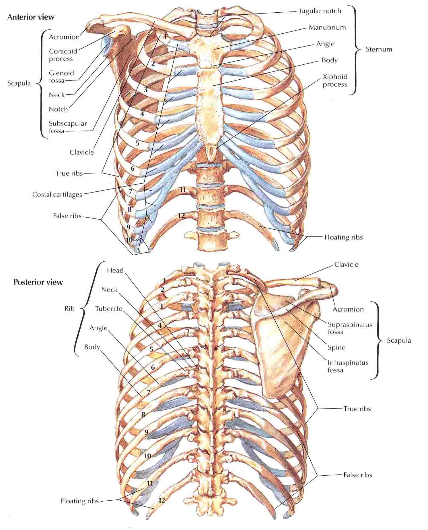
When looking at a chest X-ray it is important to remember that we are not only looking at the thorax but everything inside it too; the lungs, their markings and anatomy, the heart, boney anatomy (sternum, ribs) and soft tissue anatomy (liver). It is also important to remember that inspiration/expiration on taking a radiograph will have an affect on the appearance of it.

The Respiratory System

We should be able to see the apex/base of the lungs, the trachea and bification of such, the bronchi and the Hilum (the point at which the bronchi, blood vessels, nerves diverge from. Held together by pleura and connective tissue)

Lung markings are due to blood vessels and are important as absence of such can indicate pneumothorax and more prominent markings indicate other pathologies.


The Bronchial Tree - commences at the bification of the trachea (upper border of T5). The Right main bronchus is wider, shorter and more vertical than the left one, this means any foreign bodies may lodge in this one. The left Bronchus passes behind the arch of the aorta and in front of the oseophagus. The bronchi within  each lobe of the lung divide into smaller branches and lobules.

The right lung is separated into three lobes (Superior, medial and inferior lobe) whereas the left is only in two (<5% of people have an extra azygos lobe) Oblique (bottom) fissures and transverse (top) fissures seperate these lobes, fissures are infolding of pleural membrane which protect each lung (alongside the parietal pleura layer) These membranes contain a lubricating fluid (serous fluid) which reduces friction in respiration.  Each lung is further divided into bronchopulmonary segments composed of lobules which are wrapped in elastic connective tissue (Alveoli, Nerves, Lympathic vessels, branches of pulmonary and bronchiole arteries and terminal bronchiole).

The Heart

The heart lay inside the Mediastinum - a collection of tissues between the lungs, it is a broad partition medial to the lungs and extending from sternum and includes all contents of thoracic cavity (minus lungs).

Superior Mediastinum contains Arch of aorta
Anterior Mediastinum contains Right main pulmonary artery, left atrium, left atrial border, inferior vena cava and the right ventricle.
Middle Mediastinum contains Birfurcation of trachea and the main bronchi.
Posterior Mediastinum contains Thoracic part of decending aorta, Oesophagus, thoracic duct and lymph glands.


The ventricles of the heart have difference cardiac muscle thickness depending on how much pressure is generated (the left which supplies the whole body has a much thicker wall). Atria have comparatively little muscle wall as the pressure is a lot less there.

There are two coronary arteries which branch from the ascending aorta: the left divides into ventricular and circumflex branches. The anterior interventricular branch is in the anterior interventricular sulcus and supplies oxygenated blood to the walls of both ventricles. The circumflex lies in the coronary sulcus and distributes oxygenated blood to the walls of the left ventricle and atrium.

The right coronary artery supplies small branches to the right atrium and continues inferiorly to the right auricle dividing into posterior interventricular and marginal branches. The posterior interventricular branch supplies the walls of the two ventricles and septum with oxygenated blood. The marginal branch in the coronary sulcus transports oxygenated blood to the myocardium of the right ventricle.  

The Oseophagus

Extends from the laryngopharynx through mediastinum, diaphragm and to the superior portion of the stomach.
Composed of 4 layers 
  1. Outer areolar layer (elastic fibres - attaches it to surrounding structures)
  2. Muscular coat (muscle fibres - enables swallowing)
  3. Submucous coat (loose areolar tissue - contains vessels, nerves and muscous glands)
  4. Inner mucous coat (stratified squamous epithelium - folded rugae when empty)


Sunday, 19 February 2012

Muscoskeletal system

JOINTS

There are several types of joints:

Fibrous

These joints are fixed and move minimal amounts.

Sutures are dense fibrous connective tissue found in the skull of babies and are the 'soft spots' on their head. In older age these sutures ossify and become fixed (synostosis). The irregular interlocking edges allow for additional strength and minimise possible fractures.


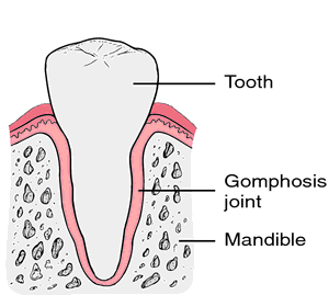
Gomphosis joints (GUMphosis) bind teeth into their bony sockets of the maxillary and mandible. The connection between tooth and socket is called the periodontal ligament.

Syndesmoses joints are found between the articulated surfaces of the tibia and fibula, made up of considerably more fibrous connective tissue than sutures and are united by interosseous ligaments.


Cartilaginous (symphysis and synchondrosis)

Cartilaginous joints have no joint cavity and are held together between cartilage, they allow more movement than in the fibrous joints but less so than synovial.

Symphysis joints such as the pubis symphysis and the external vertebral bodies are made up of broad fibrocartilage


Synchondrosis joints are made up of hyaline cartilage and can be found in epiphyseal plates, found between the first rib and the sternum. 


Synovial joints
1. Ball and socket 2. Ellipsoid joint 3. Saddle joint 4. Hinge Joint 5. Pivot Joint


These joints have a joint cavity and are subclassified by their movements, they are also usually accompanied by accessory ligaments which allow two bones of different shapes to tightly fit and stablizes the joint. Inside the joint cavity it contains synovial joint fluid which reduces friction (similar to bursae) it also supplies nutrients and removes metabolic waste and waste formed from wear and tear of cartilage by phagocytes. The articular capsule has two layers; fibrous outter layer and articulated inner layer (formed by synovial membrane). The fibrous layer allows for movement and its tensile strength reduces changes of injury and dislocation. 

Ball and socket moves on three planes; rotation, flexion/extension and abduction/adduction.


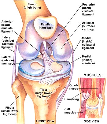
Hinge joints such as the knee consist of a convex bone surface fitting into a concave bone surface and usually only flex/extend
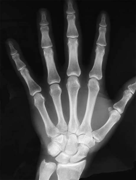
Saddle joints such as the first carpo-metacarpal joint consist of a saddle shaped articular surface and a concave/complex opposing surface. Flexion/extention, Abduction/Adduction and Circumduction.
Gliding joints such as the patellofemural are flat articulated surfaces which are restricted by ligaments so can only move side to side and back and forth.
Pivot joints can be found in the radioulnar which allow full rotation: pronation and supination.


Ellipsoidal (condyloid) joints such as the wrist joint allow two plane movement (flexion/extention, cirumduction, adduction/abduction)


Hyaline cartilage is a pearly white cartilage which is found at the ends of bones and in your ears and nose, trachea, bronchi and parts of the larynx. It provides smooth surfaces for joints to move without friction, it has a high tensile strength and is avascular so is supplied by surrounding synovial fluid.


Bursaes are fluid filled connective tissue (similar fluid to synovial fluid) which reduce friction in areas of high movement such as the knee joint.


Tendons and Ligaments

A tough band of connective tissue made of collagen fibres, they usually connect muscle to bone at periosteum; very strong and pliable. Tendons do not move at all but ligaments do to accommodate for joint movement. Tendons are inclosed by sheaths to allow them to move back and forward without friction.